Cueva de Villa Luz Microscopy and Spectroscopy of
Wall Red Goo
Click on the images to see a larger version
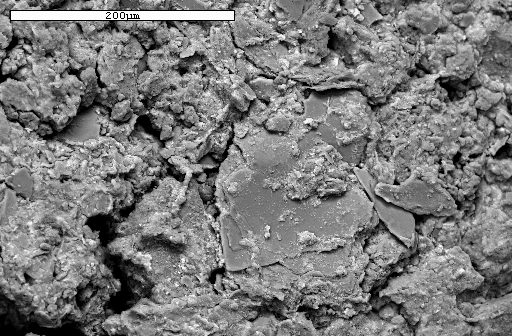
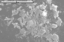
SEM image of the red clay-like substance sampled January, 1998
in the Ragu Passage. Photo by Spilde, Northup, and Boston.
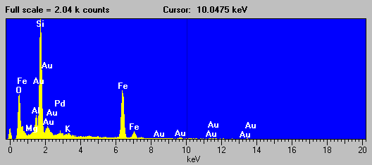
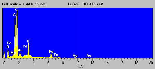
EDS spectrum from the red clay-like substance in the Ragu Passage,
showing a clay signature. Small Au and Pd peaks are from the conductive
coating applied to the sample before SEM analysis. Analysis by Spilde,
Northup, and Boston.
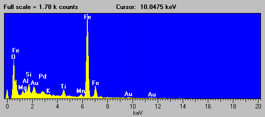
EDS spectrum from the red clay-like substance in the Ragu Passage,
showing the presence of iron oxides. Analysis by Spilde, Northup, and Boston.
EDS spectrum of the plates observed in SEM images of the red
clay-like substance in the Ragu Passage. Analysis by Spilde, Northup,
and Boston.
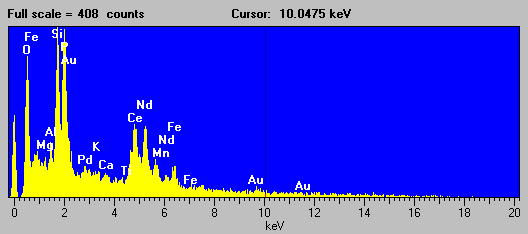
EDS spectrum of a small, Phosphorus
(P)-rich particle in the red clay-like substance sampled January, 1998
in the Ragu Passage. Note the presence of rare earths.
Analysis by Spilde, Northup, and Boston.
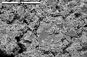
SEM image (5000x) of the red clay-like substance sampled January, 1998
in the Ragu Passage showing the presence of tiny holes.
Photo by Spilde, Northup, and Boston.
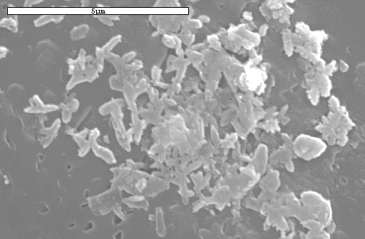
Nest of partially mineral coated bacterial shapes in the red goo.
Photo by Spilde, Northup, and Boston.
This web site Copyright 2007, 2011, Kenneth
Ingham